|
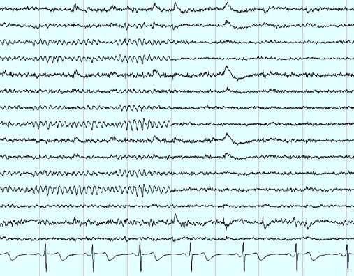
The Electro-Encephalo-Gram (EEG) is the display
of the electrical activity occurring at the surface of the brain.
The EEG is used in the evaluation of brain disorders. Most commonly it is used
to show the type and location of the activity in the brain during an Epileptic seizure.
Seizure is an uncontrolled electrical activity in the brain, which may produce a physical
convulsion or other physical signs.
This activity appears as waveforms of varying frequency and amplitude measured in voltage.
EEG waveforms are generally classified according to their frequency, amplitude, and shape, as well as the sites on the scalp at which they are recorded.
The most familiar classification uses EEG waveform frequency (e.g,. alpha, beta,
delta, theta).
Information about waveform frequency and shape is combined with the age of the patient, state of alertness or sleep, and head site to determine
clinical significance.
Normal EEG waveforms, are defined and described by their frequency, amplitude, and location. Frequency (measured
in Hertz, Hz) is a key characteristic used to define normal or abnormal EEG rhythms.
Most waves of 7.5 Hz and higher frequencies are normal findings in the EEG of an awake adult. Waves with a frequency of 7 Hz or less often are classified as abnormal in awake adults, although they normally can be seen in children or in adults who are asleep.
In certain situations, EEG waveforms of an appropriate frequency for age and state of alertness are considered abnormal because they occur at an inappropriate scalp location or demonstrate irregularities in rhythmicity or amplitude. Some waves are recognized by their shape, scalp location and symmetry.
For excellent patient education resources, visit
eMedicine's Procedures Center. Also, see
eMedicine's patient education article
Electro-Encephalo-Graphy (EEG).
|